Thoracolumbar fascia
| Thoracolumbar fascia | |
|---|---|
 Diagram of a transverse section of the posterior abdominal wall, to show the disposition of the lumbodorsal fascia. | |
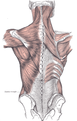
 Superficial muscles of the back. The thoracolumbar fascia is the gray area at bottom center. | |
| Details | |
| Identifiers | |
| Latin | fascia thoracolumbalis, fascia lumbodorsalis |
| TA98 | A04.3.02.501 |
| TA2 | 2242 |
| FMA | 25072 |
| Anatomical terminology | |
The thoracolumbar fascia (lumbodorsal fascia or thoracodorsal fascia) is a complex,[1]: 1137 multilayer arrangement of fascial and aponeurotic layers forming a separation between the paraspinal muscles on one side, and the muscles of the posterior abdominal wall (quadratus lumborum, and psoas major[1]: 1137 ) on the other.[2][1]: 1137 It spans the length of the back, extending between the neck superiorly and the sacrum inferiorly.[3] It entails the fasciae and aponeuroses of the latissimus dorsi muscle, serratus posterior inferior muscle, abdominal internal oblique muscle, and transverse abdominal muscle.[4]

In the lumbar region, it is known as lumbar fascia and here consists of 3 layers (posterior, middle, and anterior) enclosing two muscular compartments. In the thoracic region, it consists of a single layer (an upward extension of the posterior layer of the lumbar fascia).[3] The thoracolumbar fascia is most prominent at its lower end[1]: 814–815 where its various layers fuse into a thick composite.[2]

Anatomy
Thoracic region
In the thoracic region, the thoracolumbar fascia consists of a single layer - an upward extension of the posterior layer of the lumbar fascia, becoming progressively thinner before fading out above the 1st rib, replaced by the splenius muscle.[3]

In the thoracic region, it forms a thin fibrous fascial covering for extensor muscles associated with the spine, separating them from muscles interconnecting the spine and upper extremity.[1]: 814–815 Here it attaches to costal angles of all ribs, the spinous processes of all thoracic vertebrae, and the thoracic portion of the supraspinous ligament.[3] It is situated deep to the serratus posterior superior muscle. Superiorly, it terminates by becoming continuous with the superficial layer of deep cervical fascia of the posterior neck.[1]: 814–815

Lumbar region
The thoracolumbar fascia is most prominent inferiorly - adjacent to the caudal lumbar spine, between the posterior superior iliac spines on either side - where its aponeurotic layers meld, forming a thickened sheet.[1]: 1137 The thickened, united inferior portion attaches firmly to the posterior superior iliac spine, and the sacrotuberous ligament.[2] The thoracolumbar fascia extends as far inferiorly as the two ischial tuberotities.[1]: 1137

Function
The thoracolumbar fascia is thought to be involved in load transfer between the trunk and limb (it is tensioned by the action of the latissimus dorsi muscle, gluteus maximus muscle, and the hamstring muscles), and lifting.[1]: 814–815

It is endowed with nociceptive receptors, and may be involved in some forms of back pain.[1]: 814–815


See also
References
- ^ a b c d e f g h i j Standring S (2020). Gray's Anatomy: The Anatomical Basis of Clinical Practice (42nd ed.). New York. ISBN 978-0-7020-7707-4. OCLC 1201341621.
{{cite book}}: CS1 maint: location missing publisher (link) - ^ a b c Willard FH, Vleeming A, Schuenke MD, Danneels L, Schleip R (December 2012). "The thoracolumbar fascia: anatomy, function and clinical considerations". Journal of Anatomy. 221 (6): 507–536. doi:10.1111/j.1469-7580.2012.01511.x. PMC 3512278. PMID 22630613.
- ^ a b c d Sinnatamby C (2011). Last's Anatomy (12th ed.). Elsevier Australia. p. 274. ISBN 978-0-7295-3752-0.
- ^ "thoracolumbar fascia". TheFreeDictionary.com. Retrieved 2023-05-14.
External links
- Atlas image: back_muscles38 at the University of Michigan Health System - "Thoracolumbar Fascia, Dissection, Posterior View"
- Atlas image: back_muscles41 at the University of Michigan Health System - "Trunk, Transverse MRI Showing Lamellae of the Thoracolumbar Fascia"
See what we do next...
OR
By submitting your email or phone number, you're giving mschf permission to send you email and/or recurring marketing texts. Data rates may apply. Text stop to cancel, help for help.
Success: You're subscribed now !
